EN
தமிழ்
KM NU Hospitals accreditation

Hip Pain: Painless and Surgical Treatment Options

Hip pain is a common symptom that can be brought on by anything from arthritis to sports injuries. It can interfere with daily routine and exercise, making even simple tasks such as walking or bending seem difficult.

Hip pain is typically treated at home with over-the-counter painkillers and taking a break from physical activity. However, additional therapies might be required if the pain worsens or doesn’t go away.

Reasons for hip pain

Part of the treatment plan for hip main includes finding out the cause of hip pain. Some common causes include:

·       Bursitis: Bursae are sacs of fluid found between the bones and soft tissues which reduce friction during movement. Bursitis is inflammation of the bursae, resulting in pain on the outer hip.

·       Hip strain: When a hip muscle is overstretched or tears, it can weaken the joint and cause pain and swelling.

·       Tendinitis: Tendons are the soft tissues that connect muscles with bones. Inflammation of tendons causes pain during movement.

·       Osteoarthritis: This is a common form of arthritis in which the cartilage in the hip joint breaks down.  It may remove the cushioning between bones, resulting in hip stiffness, decreased range of motion, and pain in the groin and thighs.

·       Rheumatoid arthritis: This type of arthritis is an autoimmune disorder in which the body’s immune system causes cartilage-destroying inflammation throughout the body, especially in the hips. Rheumatoid arthritis causes discomfort that worsens with rest but improves with activity.

Treatment options for hip pain

Treatment modality for hip pain may be classified as non-surgical and surgical techniques. Here, we describe each of these techniques in detail.

A.     Non-surgical hip pain treatment

(i) Injections and infusions: Corticosteroid injections can be directly administered into the hip to relieve pain and inflammation. In infusion therapy, medications are directly delivered into the muscles or intravenously in order to reduce inflammation. 

The latest technique in the form of PRP or platelet-rich plasma injections make use of platelets from your own blood which boosts tissue regeneration, aids in wound healing, and repairs cartilage.

(ii)  Nutraceuticals: These are herbal or dietary supplements that help reduce pain in the hip joint. Examples of such supplements are glucosamine and chondroitin, which may help strengthen overall health of the bones and soft tissues.

(iii) Lifestyle modifications: Losing the excess flab, indulging in low-impact exercises, and using a cane or walker are some simple lifestyle modifications that can be incorporated to decrease hip pain.

(iv)Pain medications: Over-the-counter (OTC) pain relievers can be used to ease hip pain. Analgesics such as muscle rubs can also bring temporary pain relief. Other than these, prescription medications such as antirheumatic drugs and biological response modifiers may also be used.

(v)Physical and occupational therapy: Physical therapy is especially beneficial if you suffer from arthritis. As part of the exercise programs, your physical therapist will teach you some of the safer forms of exercise that will benefit your hip. You will also be taught ways to complete daily activities with less discomfort.

(vi)  RICE method: RICE is an acronym for “rest, ice, compression, and elevation.” This comprises of taking rest from pain-inducing activities, applying ice pack over the painful area, wearing a compression bandage on the hip to facilitate blood flow, and keeping the affected leg at an elevated height, above the level of the heart while resting.

B.      Surgical hip pain treatment: When all the above non-surgical hip pain treatment methods fail, surgical options may be your best bet.

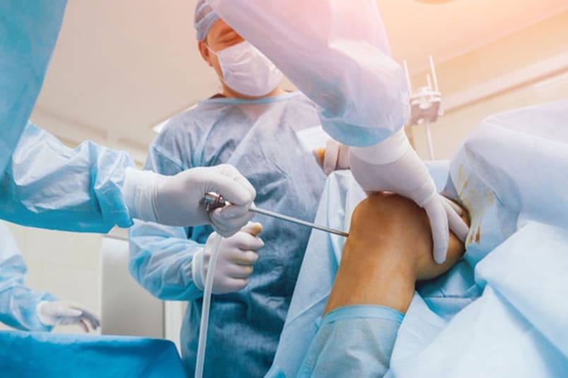
(i)  Hip arthroscopy: In order to remove or repair damaged soft tissues or remove problematic bone fragments, the surgeon will make a few small incisions in the hip and use specialized surgical instruments and a scope equipped with a camera.

(ii)  Hip replacement (Hip arthroplasty): In this procedure, an orthopaedic surgeon replaces one or both of the damaged hip joint’s ends with artificial components. The damaged sections of the hip joint are replaced with parts that are made up of metal, ceramic, or very hard plastic.

To summarize briefly, hip pain is a common symptom that can occur due to sports injury, a medical condition, or because of accidents and falls. With a host of treatments on offer these days, thankfully there are more than one alternative. 

To establish which treatment method would be most suitable for your particular situation, it is advisable to get in touch with your healthcare provider who would decide on the right course of treatment. 

The aim of the treatment resorted to should be to reduce pain and inflammation while allowing the individual the liberty to carry out daily chores with less restriction and limitations.

KM NU Hospitals is a renowned name in the field of orthopedic care.  Located in Ambur, Tamil Nadu, this institute is famous for its expert team of medical professionals, state-of-the-art facilities, and top-notch medical equipment. 

The orthopedic surgeons at KM NU Hospitals are adept and experienced in handling numerous hip replacement surgeries successfully. If you are suffering from hip pain and want a fool-proof solution for it, visit KM NU Hospitals in Ambur to avail the best in-house treatment for hip pain.

References:

1.  Hip pain. Cleveland Clinic. https://my.clevelandclinic.org/health/symptoms/21118-hip-pain.

2.  Hip pain treatments: What are your options? Reid Health. https://www.reidhealth.org/blog/hip-pain-treatments-what-are-your-options.

3.  Non-surgical hip pain treatment. Penn Medicine. https://www.pennmedicine.org/for-patients-and-visitors/find-a-program-or-service/orthopaedics/hip-pain/non-surgical-hip-treatment#:~:text=Pain%20Medications%3A%20Over%2Dthe%2D,used%20for%20temporary%20pain%20relief.

4.  Treatment options for hip pain. HSS. https://www.hss.edu/hip-pain-center-treatments.asp.

Author: Dr. Allen Sunny Deol S V

6 Simple Exercises To Improve Mobility After Hip Replacement

Mobility is defined as the ability of a person to move about, which includes actions such as standing up and walking. After a hip replacement or hip arthroplasty surgery, getting back to doing normal chores and activities requires a person to be mobile once again.

The strategies taken to help a person become mobile again post a hip replacement surgery fall under mobility strategies. Here, we cover a few of the mobility strategies that can help you get on your feet once more after hip replacement surgery.

Hip replacement surgery


In this surgery, the ball and socket part of the hip joint is replaced. A metal implant (stem) and another metal and plastic implant (cup) is inserted in the thigh bone and pelvic bone, respectively.

There is a metal head which moves in the plastic cup, relieving pain and improving mobility. Almost 90% of the hip replacement surgeries are performed on older adults above 50 years.

There are two types of hip replacement surgeries:

(i)  Total hip replacement: This is the most common form of hip replacement in which the whole hip is replaced with a prosthetic joint. In this surgery, femoral head (top) of your thigh bone (femur) and the socket it fits into, called acetabulum is replaced.

(ii)  Partial hip replacement: This type of surgery is very rare, in which only a part of your hip gets replaced. These are only done to remove certain types of tumors and hip fractures (broken hips).

Resuming normal life


Resuming normal activities after a hip replacement surgery may seem like a daunting task for many, especially after remaining immobile for long.

To overcome this barrier after initial bed rest and restricted weight bearing, you may require professional assistance.

There are various strategies to improve mobility, including resuming walking and exercise programs used during your stay in the hospital and even after discharge.

As part of effective treatment for improving people’s mobility, exercises in addition to standard physiotherapy is recommended. These exercises focus on balance, walking, and functional tasks while you remain in the hospital. Once discharged from the hospital, the target should be on extra strength and endurance training

Exercises for your hip

Your surgeon or physical therapist will put you on an exercise and stretches regime as early as 24 hours after the surgery has completed. You will be closely monitored to assess strength and flexibility in your legs and hip and how long you are able to stand and sit.

The exercises you need to follow are centered around strengthening and stretching the muscles around your hip joint. You will be able to slowly resume activities such as climbing stairs, bending, and walking.

The following hip exercises are beneficial after hip replacement surgery:

1.  Straight leg raises: Lie on your back. Now, keeping your legs straight, lift alternate legs 6 inches off the floor. Hold till the count of 5 in this position and relax. Repeat 20 times.

2.  Hip abduction: Lie on your back, keeping toes pointed toward the ceiling. Now move one leg out to the side as far as possible. Slowly return to the starting position and relax. Repeat with the other leg.

3.  Bridging: Lie on your back, bending both knees. Push feet and lift your back. Hold in this position for a count of 5 and then relax. Repeat.

4.  Hip adduction: Lie on your side, with the painful side of the hip up. Keeping the top leg straight, lift it as high as possible. Hold till the count of 5 and then relax. Repeat again.

5.  Hip extension: Lie on your stomach and lift your leg up by putting pressure on the thigh. Hold for a count of 5 and bring the leg down. Relax and repeat with alternate leg.

6.  Isometric glutes: Squeeze or contract your buttocks while lying on your stomach. Hold till the count of 5 and relax. Repeat 10 times.

With the help of the above exercises, you will regain your range of motion and strength over time. It may be nearly 6-12 weeks before you can use your hips without any restrictions.

Although you need time to recover after a hip replacement surgery, with proper medical guidance and exercise plan, you will be able to resume daily activities soon. You may have to use a cane or crutches to help you in walking till the time proper balance is restored to avoid unnecessary trips and falls

Every individual has a varying recovery period which is subject to the exercise regime followed. With diligence and care, you will soon find yourself back on your feet!

At KM NU Hospitals, which is the best hip fracture surgery hospital in Ambur, Tamil Nadu, you will find yourself surrounded by orthopaedics specialists who are experts in hip fracture treatment. This healthcare facility offers the latest techniques for hip arthritis surgery in Ambur. If you need advice regarding your hip replacement surgery or want to undergo the same, KM NU Hospitals is the right place for you.


References:

1.  Hip Replacement (Hip Arthroplasty). Cleveland Clinic. https://my.clevelandclinic.org/health/treatments/17102-hip-replacement.

2.  Total Hip Replacement Exercise Guide. OrthoInfo. https://orthoinfo.aaos.org/en/recovery/total-hip-replacement-exercise-guide/#:~:text=Proper%20walking%20is%20the%20best,regain%20movement%20in%20your%20hip.

3.  Your all-you-need-to-know guide on hip replacement surgery. TOPDOCTORS UNITED KINGDOM. https://www.topdoctors.co.uk/medical-articles/your-all-you-need-to-know-guide-on-hip-replacement-surgery.

Author: Dr. Allen Sunny Deol.S.V

C-Section Pregnancy Scars: Prevention, Management, Causes & Types

A Caesarean section, commonly known as a C-section, is a surgical procedure for delivering a baby through an incision in the mother’s abdomen and uterus. This method is employed when a natural birth is risky for the mother, baby, or both. 

C-sections may be planned or performed in emergencies, addressing complications such as abnormal positioning, foetal distress, or maternal health concerns. The procedure involves anaesthesia, an incision, and careful removal of the baby. 

While C-sections are generally safe, they entail recovery time and potential risks. The decision to undergo a C-section is typically made collaboratively by healthcare providers and expectant parents.


Types of caesarean sections

The choice of the type of caesarean section depends on various factors, including the reason for the surgery, the position of the baby, and the mother’s medical history. It is performed to ensure a safe delivery while minimizing risks to both the mother and the baby. The following are the commonly used C-section incisions:


. Low transverse caesarean section: This is the most common type of C-section. The incision is made horizontally across the lower part of the uterus, also known as a “bikini cut.” It is preferred over the classical incision because it is associated with less bleeding and a lower risk of uterine rupture in future pregnancies.


. Classical incision:  This type involves a vertical incision made on the upper part of the uterus. It is rarely performed nowadays, due to its association with a higher risk of uterine rupture in subsequent pregnancies.


Causes of C-Section Scars


The C-section incision typically results in a scar, and various factors can contribute to the formation and appearance of scars. The following are some common causes:

. Surgical technique: The way the incision is made and closed during the C-section can affect the appearance of the scar. Proper surgical technique, including precise closure of the incision, can help minimize scarring.

. Infection: Infections that occur during or after the C-section can contribute to the formation of noticeable scars. Infections can hinder the natural healing process and may result in a thicker or more raised scar.

. Poor wound care: Inadequate care of the incision site can lead to complications such as infection or delayed healing, which may affect the final appearance of the scar. Following postoperative care instructions from healthcare providers is important.

. Genetics: Some individuals are more prone to developing noticeable scars due to genetic factors. If a person has a family history of hypertrophic or keloid scarring, they may be more likely to develop such scars after a C-section.

. Obesity: Women with higher body mass index (BMI) may have an increased risk of developing larger and more prominent scars after a C-section.

. Diabetes: Diabetes can impact the body’s healing process, potentially leading to slower wound healing and an increased risk of complications, including poor scar formation.

. Pregnancy complications: Complications during pregnancy or labour that necessitate an emergency C-section may contribute to a higher likelihood of scarring. 

.  Previous abdominal surgeries: If a woman has had prior abdominal surgeries, especially those involving the uterus, there may be an increased risk of adhesions and scar tissue formation during subsequent C-sections.


Management of C-Section Scars

Management of C-section scars involves various options to reduce their visibility and improve skin appearance: The following are the various treatment options for C-section scars:

·       Silicone treatment: Utilizing silicone gel or sheets/tapes has proven effective in minimizing surgical scars, particularly for smaller scars. The application of silicone aids in breaking down scar tissue, leading to a lighter and less noticeable scar over time.

·       Steroid injections: For larger scars, steroid injections are employed to flatten the affected area, making the scar less conspicuous. Regular visits to a cosmetic gynaecologist are necessary for continued injections, as it is not a one-time procedure.

·       Laser therapy: Advanced medical lasers can be used to lighten C-section scars, even those that are a few months old. Multiple sessions may be required to achieve visible results, making this an effective option for scar reduction.

·       Scar revision surgery: In cases where other treatments prove insufficient, scar revision surgery may be recommended. This procedure involves making an incision over the scar, removing the scar tissue, and allowing the body to heal anew. If the production of scar tissue is controlled, the visibility of the scar can be reduced. Subsequent treatments may then be explored to further lighten the scar.


Prevention of C-Section Scars

Preventing or minimizing the appearance of C-section scars involves proper wound care and promoting overall skin health. While it is essential to note that individual healing varies, below are some tips to help prevent or reduce the visibility of C-section scars:


·       Follow medical advice: Adhere to your healthcare provider’s instructions for post-operative care. This includes keeping the incision area clean, dry, and protected to prevent infection.

·       Gentle cleansing: Clean the incision site with mild soap and water as recommended by your healthcare provider. Avoid chemicals, scented products, or excessive scrubbing.

·       Keep it dry: Ensure the incision area remains dry. Rub it gently with a clean, soft towel after washing.

·       Avoid sun exposure: Protect the scar from direct sunlight, especially during the initial healing period. Use sunscreen (SPF 30 or higher) or cover the scar with clothing when exposed to the sun to prevent hyperpigmentation.

·       Stay hydrated and eat a healthy diet: Proper nutrition is essential for better skin health and faster wound healing. Stay hydrated and consume a balanced diet rich in vitamins and minerals, especially vitamin C and zinc.

·       Manage your weight: Avoid rapid weight gain or loss, as fluctuations can impact the appearance of scars.

·       Massage: Gentle massage of the scar tissue can promote blood circulation and help reduce scar thickness.  

·       Scar creams and ointments: Some over-the-counter scar creams or ointments containing ingredients like silicone, vitamin E, or onion extract may help reduce scarring. Consult your healthcare provider before using any products to ensure they are safe for your specific situation.

·       Stay active: Gentle physical activity can help improve blood circulation and promote overall healing. However, avoid strenuous exercises until you receive instructions from your healthcare provider.


To summarize, caesarean section scars require careful postoperative care to minimize complications. Adequate wound hygiene, monitoring infections, and avoiding strenuous activities during the initial healing phase are essential. Regular follow-ups with healthcare professionals ensure proper assessment and timely intervention if issues arise. Adhering to medical advice promotes optimal healing and reduces the risk of complications, fostering a successful recovery for mothers with caesarean section scars.

NU Fertility in Bangalore, India, is renowned for delivering exceptional cosmetic gynaecological services, particularly in caesarean sections. With a commitment to precision and patient care, NU Fertility stands out for its expertise in enhancing the aesthetic aspects of caesarean procedures, ensuring both medical excellence and personalized attention. Experience the best cosmetic gynaecological services at NU Fertility, where your well-being and satisfaction are prioritized with utmost care.


References:

1.  Scar Tissue after a Cesarean Section. National Library of Medicine.

https://www.ncbi.nlm.nih.gov/pmc/articles/PMC8620716/.

2.  C-section. Medline Plus.

https://medlineplus.gov/ency/patientinstructions/000624.htm.

3.  C-section. Better Health.

https://www.betterhealth.vic.gov.au/health/healthyliving/caesarean-section


Author: Dr. Gracy Sunny Kindo

Preterm Labor: Symptoms, Causes, & Risk Factors

Having a smooth and full-term pregnancy, without any complications is the dream of every woman who thinks of attaining motherhood. For this, a lot of medical check-ups, including blood tests and imaging tests are regularly done to monitor the progress of the foetus. However, despite taking all necessary precautions, there are times when the woman experiences preterm labor. 

Preterm labor is said to occur when regular contractions take place around week 20 instead of week 37, which results in the opening of the cervix. A baby born as a result of preterm labor faces greater health risks and may require to be kept under observation in the neonatal intensive care unit.


What is premature birth?

Premature birth or preterm birth, as it is also known, refers to when a baby is born before 37 weeks of gestational period. Preterm babies are often seen to suffer from health complications such as trouble in breathing, underdeveloped organs, vision problems, and low birth weight.


Signs and symptoms of preterm labor


There are many signs and symptoms related to preterm labor which can easily be recognized. Some of these indications include:

·       Frequent or regular sensations of abdominal tightening (contractions): This means experiencing four or more contractions in one hour that don’t go away even after changing your position or relaxing.

·       Sensation of pelvic or lower abdominal pressure: Regular low, dull pain in your back that either comes and goes on its own or is constantly present and can’t be relieved by changing positions or by using other comfort measures.

·       Mild abdominal cramps: These cramps are accompanied with or without diarrhoea that sometimes might feel like gas pain.

·       Continuous low, dull headache: Headache is a common complaint in women who experience preterm labor.

·       Vaginal spotting or light bleeding: Sometimes, the woman might exhibit persistent menstrual-like symptoms, including spotting or light bleeding.

·       Preterm rupture of membranes: This may take place in a gush or as a continuous trickle of fluid, after the membrane around the foetus breaks or tears.

·       Watery, mucus-like, or bloody vaginal discharge: There may be a change in the type of vaginal discharge. It may also be more than usual.

·       Nausea and vomiting: You may also experience flu-like symptoms such as nausea and vomiting.

·       Decreased foetal movements: If you don’t feel the baby make at least six movements in the span of an hour, it might be an indication of preterm labor.


Causes of preterm labor

You may experience premature or preterm labor if you:

·       Had a premature baby in the past

·       Have a pregnancy with twins, triplets, or more

·       Have or had a problem in the past with your cervix, uterus, or vagina


Risk factors for preterm labor

There are few risk factors associated with preterm labor. They may be divided into the following four classes:

A.     Pregnancy risk factors

·       Vaginal bleeding throughout the pregnancy

·       Pregnancy achieved via in vitro fertilization (IVF)

·       Carrying a foetus suspected to have a congenital abnormality

·       Short spacing between two pregnancies


B.      Age and race risk factors

·       Age less than 17 or greater than 35 years

· Belong to the Black race


C.      Medical risk factors

· Untreated vaginal infections such as urinary tract infections (UTIs), sexually transmitted infections (STIs), etc.

·       Placental problems such as placental abruption

·       Connective tissue problems such as Ehlers-Danlos syndrome (EDS)

·       Excess of amniotic fluid (polyhydramnios) or too little amniotic fluid (oligohydramnios)

·       Short cervix or irregularly-shaped uterus


D.      Lifestyle risk factors

. Lacking prenatal care

. Being underweight or overweight before pregnancy

. Not gaining enough weight during pregnancy

. Consuming a poor diet

. Smoking, drinking, or using illegal drugs

. Demanding job that requires long hours of standing

. Having high stress or anxiety levels

. Being exposed to teratogens such as chemicals, lead, radiations,

and other harmful substances

Having seen the symptoms, causes, and risk factors for preterm labor, it is important to ensure that you are aware of these. Even though it may sound difficult, try not to panic as the symptoms usually wear out on their own and most women give birth at term. 

Preterm labor is usually temporary. In case you do go into preterm labor during your pregnancy, notify your healthcare provider immediately who will recommend certain medications to stop or delay the labor. 

Premature labor can be prevented with bed rest, intravenous fluids, and uterine relaxants (medications). Preterm labor can be avoided most effectively by maintaining good health throughout your pregnancy and taking care of any pre-existing health conditions.

KM NU Hospitals is located in Ambur, Tamil Nadu. It has the best gynecologist and obstetrician in Ambur who are well-qualified to deal with pregnancy-related emergencies such as preterm labor. 

KM NU Hospitals is known for its state-of-the-art facilities and highly advanced technology which has helped bring a smile to millions of people. Visit this premiere medical institute today without delay!

Lower Respiratory Tract Infection in Children: Prevention and Treatment Guide | KM NU Hospitals

Lower Respiratory Tract Infections (LRTIs) in children refer to infections that affect the airways and lungs. These conditions can include various illnesses, such as pneumonia and bronchiolitis.

LRTIs are typically caused by viral or bacterial pathogens, leading to various symptoms. Timely diagnosis and appropriate management are crucial for minimizing the impact on respiratory function and preventing long-term consequences. 

This blog aims to provide various insights about common LRTIs in children, their treatment, and prevention strategies.

Common LRTIs in Children

LRTIs in children are common and can be caused by various infectious agents. The following are the common lower respiratory tract infections in children:

Bronchiolitis: This is a common viral infection in infants and young children, often caused by the respiratory syncytial virus (RSV). It primarily affects the small airways (bronchioles) in the lungs and can lead to symptoms such as coughing, wheezing, and difficulty breathing.

Pneumonia: An infection that causes the inflammation of the air sacs in one or both lungs, caused by bacteria, viruses, or other microorganisms. Common pathogens include Streptococcus pneumoniae, Haemophilus influenzae, and viruses such as influenza and adenovirus.

Influenza (Flu): A viral infection that can affect the upper and lower respiratory tract. It typically shows symptoms such as fever, cough, sore throat, body aches, and fatigue.

Pertussis (Whooping Cough): It is a bacterial infection caused by Bordetella pertussis. It can result in severe coughing fits, especially in young children, and may lead to complications.

Symptoms of LRTIs in Children

The symptoms of LRTIs can vary in severity, and not all children will experience the same set of symptoms. Common signs of lower respiratory tract infections in children include:

·   Cough

·   Wheezing

·   Difficulty breathing

·   Chest pain

·   Fever

·   Fatigue

·   Nasal congestion

·   Cyanosis (bluish discoloration of skin and lips)

Diagnosis of LRTIs in Children

The following are the various exams and tests that can be used to diagnose lower respiratory tract infections (LRTIs) in children:

Medical history and physical exam: This is the initial step where the doctor gathers information about the child’s symptoms, previous illnesses, and overall health. The physical exam helps identify specific signs and symptoms related to LRTIs.

Nose swab: Swabbing the nose helps identify the presence of viruses such as the flu. Since antibiotics are ineffective against viral infections, this test helps guide treatment decisions.

X-ray: If pneumonia is suspected, an X-ray of the chest can provide a clear image of the lungs. Pneumonia often presents as an area of increased opacity on the X-ray due to inflammation and fluid in the lungs.

Blood test: Blood tests can reveal signs of inflammation or an ongoing infection. Elevated white blood cell count is a common indicator that the body is responding to an infection.

Bronchoscopy: This is a more invasive procedure, typically used for cases with chronic health conditions or recurrent pneumonia. It involves inserting a flexible tube into the airways to examine and collect samples. It allows for a direct visualization of the lower airways and helps identify the cause of the infection.


Treatment of LRTIs in Children

The treatment of Lower Respiratory Tract Infections (LRTIs) in children depends on the specific cause of the infection. LRTIs can be caused by viruses or bacteria, and the treatment approach may vary accordingly. It is important to note that many respiratory infections in children are viral, and antibiotics are not effective against viruses. Below are general guidelines for the treatment of LRTIs in children:

Antibiotics:  Antibiotics are commonly prescribed for bacterial LRTIs in children. Conditions such as bacterial pneumonia or bronchitis may necessitate antibiotics to target the specific causative bacteria.

Supportive care: Supportive care is important in managing LRTIs. Adequate hydration, maintaining a comfortable environment, and using a humidifier, can help ease symptoms and promote recovery. Over-the-counter medications may be used under medical guidance.

Bronchodilators: For cases of bronchiolitis or asthma-related LRTIs, bronchodilators may be employed. These medications help relax the airway muscles, facilitating better airflow.

Corticosteroids: In some cases, corticosteroids may be used to reduce inflammation and alleviate symptoms. However, their use is usually reserved for specific cases under medical supervision.

Hospitalization: Severe LRTIs may require hospitalization, especially if a child experiences difficulty breathing, persistent high fever, or dehydration. Hospital-based care allows close monitoring and intervention, if necessary.

Prevention of LRTIs in Children

Preventing LRTIs in children involves a combination of strategies, many of which focus on reducing the risk of exposure and strengthening the child’s immune system. Here are some preventive measures:

Vaccination: Ensure that your child receives all recommended vaccines, including those for influenza and pneumonia. Vaccination is one of the most effective ways to prevent certain respiratory infections.

Good hygiene practices: Encourage regular handwashing with soap and water, especially after coughing, sneezing, or being in contact with someone who is sick. Teach children to avoid touching their faces, especially their eyes, nose, and mouth, with unwashed hands.

Respiratory hygiene: Educate and reinforce proper cough and sneeze techniques. Encourage covering the mouth and nose with a tissue or the elbow, rather than the hands. Dispose off the used tissues properly and wash hands immediately afterwards.

Avoid exposure to tobacco smoke: Avoid exposing children to second-hand smoke, as it can significantly increase the risk of respiratory infections. If you smoke, do so away from children, and consider quitting the habit.

Maintain a healthy environment: Keep living spaces clean and well-ventilated. Regularly clean and dust the house to minimize potential irritants. Use a humidifier in dry climates, as maintaining adequate humidity can help prevent respiratory infections.

Proper nutrition: Ensure that your child has a balanced and nutritious diet to support a healthy immune system. Incorporate a diverse range of fruits, vegetables, whole grains, and protein-rich foods.

Adequate Sleep: Make sure that your child gets enough sleep. An adequately rested body is more proficient at defending against infections.

 Regular Exercise: Encourage regular physical activity to support overall health and strengthen the immune system.

Limit exposure to sick individuals: Avoid close contact with individuals who are sick, and keep sick children at home to prevent the spread of infections.

Stay informed: Being aware about local outbreaks of respiratory infections. Follow public health guidelines and seek medical advice promptly if your child shows symptoms of a respiratory infection.

To summarize, LRTIs pose a significant health threat to children, causing morbidity and mortality worldwide. If you suspect your child has a respiratory infection, it is essential to seek medical attention immediately. Early intervention and appropriate treatment can help prevent complications and promote a quicker recovery. Additionally, make sure your child is up-to-date on vaccinations, as many vaccines protect against common pathogens that can cause LRTIs.

KM NU Hospitals in Ambur, India, excels in treating Lower Respiratory Tract Infections in children, offering state-of-the-art medical care, provided by a dedicated team of experts. Their focus extends beyond treatment to encompass effective preventive measures, ensuring the well-being of young patients. With a commitment to excellence, KM NU Hospitals stands out as a reliable healthcare destination for addressing LRTIs in children, combining advanced medical interventions with a proactive approach to prevent respiratory infections.

References:

1.Lower Respiratory Tract Infection. Riley Children’s Health. https://www.rileychildrens.org/healthinfo/lowerrespiratorytractinfection.

2.Lower Respiratory Tract Infection. Patient. https://patient.info/doctor/lowerrespiratorytractinfectioninchildren.

3.Viral Lower Respiratory Tract Infection. National Library of Medicine. https://www.ncbi.nlm.nih.gov/pmc/articles/PMC1126381/

Author: Dr. Palani Rajan P

Ways to Manage Knee and Joint Pain

Pain can be very debilitating, especially if it restricts your body movements. Having knee or joint pain can be extremely bothersome, particularly if you are suffering from arthritis. Knee and joint pain can be treated at home by taking enough rest and using over-the-counter medications. It is only when the pain interferes with normal activities and continues for a couple of days in a row, that one should consult a healthcare provider.

What is knee pain?

Any pain or discomfort that you experience in and around the knee joint qualifies as knee pain. Joints are places where bones are connected to each other. The knee joint is the place where the thigh bone (femur) meets the shin bone (tibia). The knee joint is the largest joint in the body and bears the whole weight of the body. Knee pain can be temporary, short-term, or chronic (long-term) in nature. You might experience pain:

·       Close to the surface, above, or behind the knees

·       Deep inside the knees

·       In multiple areas of the knees and legs

Common causes of knee or joint pain

The three most common causes of knee pain include:

1.  Overuse: Strenuous physical activities such as exercise, playing sports, and doing physical work can cause wear and tear of the knee joint. The pain gets worse when we do the same repetitive motion like working on your hands and knees and jumping a lot. Conditions arising due to overuse:

·       Bursitis

·       Osgood-Schlatter disease (jumper’s knee)

·       Patellofemoral pain (runner’s knee)

·       Tendinitis


2.  Arthritis: Arthritis results in pain and inflammation of the joints. Knee arthritis is a common occurrence. The symptoms related to knee arthritis are swelling, pain, and stiffness. Several forms of arthritis that can cause knee pain include:

·       Osteoarthritis

·       Rheumatoid arthritis

·       Post-traumatic arthritis


3.  Injuries: Any injury to the knee joint can cause pain. Some common knee injuries could be related to:

·       Sports injuries

·       Repetitive strain injuries

·       Traumas such as falls or car accidents

Knee pain may also be caused due to injuries that arise due to knee sprains, hyperextended knees, knee ligament tears, meniscus tears, bone fractures, and dislocations.

Knee and joint pain treatments

Depending on the cause and severity of pain, a treatment plan may be charted by your healthcare provider.

A.     RICE method for knee pain- This is a home-based treatment method which stands for rest, ice, compression, and elevation.

(i)              Rest: Discontinue the physical activity that has caused the knee pain in the first place so as to avoid making the injury worse.

(ii)                Ice: On the first day after the injury, apply ice pack or use a cold compress for 15 to 20 minutes every hour. After one day, apply ice every 3-4 hours. However, care should be taken not to apply ice directly but to wrap it in a towel or washcloth first.

(iii)              Compression: Compression lessens swelling and blood flow to your injured knee. Wrap a compression bandage around your knee.

(iv)              Elevation: Prop your knees using a pillow or cushion to bring it above the level of your heart.


B.      Medications: Over-the-counter non-steroidal anti-inflammatory drugs (NSAIDs) may be helpful to alleviate knee pain and other associated symptoms. A word of caution here–

do not take these medications for more than 10 days without consulting your healthcare provider or if you suffer from kidney or liver disease.


C.      Knee braces: Your knee is held in place and supported by a knee brace. Knee braces function by maintaining the alignment of your knee. Usually composed of metal or stiff plastic, they have straps that go around your leg and knee. Consult your healthcare provider regarding the type of brace you will need and how often you should wear it.


D.     Physical therapy: Physical therapy is helpful if you are suffering from arthritis or are recovering from a knee injury. Your therapist will teach you stretches and exercises that will strengthen the muscles around your affected knee. This helps in bringing relief in pain.


E.       Knee surgery: A knee surgery is generally required if you are suffering from severe arthritis, a damaged ligament, or bone fracture. When no other treatment options work and the pain is severe, a surgery is called for. Knee arthroscopy is the most common form of knee surgery where a special tool called an arthroscope is inserted into the knee joint, after making a few incisions. The arthroscope comes with a fitted camera and light that allows the surgeon to see and repair the damaged knee.

Sometimes, you may need to undergo a knee replacement (arthroplasty) which means that the surgeon replaces the knee joint with an artificial implant (prosthesis). A knee replacement is suggested in case the pain in the knee affects your ability to stand, walk, and move. 

Prevention of knee pain

Although neither injuries can be completely avoided nor can arthritis be prevented, yet some measures can be taken to mitigate these from occurring. The ideal way to prevent pain is to stay safe while being active. Ensure that you do the following while engaging in sports and other physical activities:

·       Wear the right protective gear.

·       Give your body time to rest and recover after intense physical activity.

·       Warm up or stretch before playing sports or exercising.

·       Cool down and stretch after each physical activity.

For reducing the risk of injuries due to falls, practice the following safety tips:

·       Make your home and workplace clutter-free to reduce the chances of trips and falls.

·       Use proper tools to reach for things. Do not stand on chairs, tables, and countertops.

·       Support yourself with a cane or walker if you have difficulty walking or are more prone to falls.


Everyone suffers from knee and joint pain at some point of time in their lives. While diseases such as arthritis cannot be prevented, you can reduce the risk of falls and trips by following the above guidelines. Ensuring safety both at home and in the workplace will keep you away from injuries that lead to knee and joint pain. In case you do inflict some injury upon your knees or there is pain due to arthritis, consult your healthcare provider on the treatment options available for you.

KM NU Hospitals at Ambur is one of the top multispecialty hospitals in Tamil Nadu. With a team of passionate and dedicated doctors, this healthcare set up is renowned for its orthopedic treatments. If you or a loved one is looking for a knee replacement treatment, this is the best place to opt for your treatment. Visit KM NU Hospitals without delay and get yourself relieved from knee and joint pain.


References:

1.  Knee pain. American Society of Anesthesiologists. https://www.asahq.org/madeforthismoment/pain-management/types-of-pain/knee-pain/.

2.  Knee pain. Cleveland Clinic. https://my.clevelandclinic.org/health/symptoms/21207-knee-pain.

3.  Knee pain. Mayo Clinic. https://www.mayoclinic.org/diseases-conditions/knee-pain/diagnosis-treatment/drc-20350855.

4.  Take control of your knee pain. Harvard Medical School. https://www.health.harvard.edu/pain/take-control-of-your-knee-pain.

5.  What is knee pain? VERSUS ARTHRITIS. https://versusarthritis.org/about-arthritis/conditions/knee-pain/.

Author: Dr. Tahir Ahmed

Complete Guide to a Healthy Pregnancy

Pregnancy is a transformative and exciting journey that requires careful attention to both physical and emotional changes. Ensuring a healthy pregnancy is crucial for the well-being of both the mother and the developing baby. This blog aims to provide comprehensive insights into maintaining a healthy pregnancy, covering various aspects from nutrition to exercise and psychological health.

Guide to a Healthy Pregnancy

Maintaining a healthy pregnancy involves a holistic approach encompassing physical, emotional, and lifestyle factors.  The following are the various aspects to be considered for overall maternal and foetal health:

Preconception health:

Before conception, ensure optimal health by choosing a balanced diet, exercising regularly, and managing stress. Consult a healthcare provider to address any pre-existing medical conditions and ensure vaccinations are up to date.

Early prenatal care:

Schedule an early prenatal check-up to confirm the pregnancy and receive essential guidance. Begin taking prenatal supplements containing folic acid to reduce the risk of neural tube defects in the developing baby.

Nutrition:

Adopt a well-rounded, nutrient-rich diet. Consume a variety of fruits, vegetables, whole grains, lean proteins, and dairy products. Stay hydrated and limit caffeine intake. Consult a healthcare provider to determine appropriate weight gain.

Exercise:

Engage in moderate-intensity exercise, such as walking or swimming, unless advised otherwise by a healthcare professional. Regular physical activity can enhance mood, reduce discomfort, and promote overall well-being.

Avoid harmful substances:

Eliminate alcohol, tobacco, and recreational drugs to safeguard the baby’s development. Minimize exposure to environmental toxins, and consult healthcare providers before taking any medications or supplements.

Regular prenatal check-ups:

Attend scheduled prenatal appointments to monitor the baby’s growth and address any concerns. Routine screenings and ultrasounds help detect potential issues early, allowing for timely intervention.

Educate yourself:

Stay informed about pregnancy, childbirth, and postpartum care. Attend prenatal classes, read reputable sources, and discuss any questions or concerns with healthcare providers.

Emotional well-being:

Pregnancy brings emotional challenges. Prioritize mental health through relaxation techniques, meditation, and connecting with supportive friends and family. Seek professional help if needed, as maintaining emotional well-being is crucial for both the mother and the baby.

Sleep:

Ensure adequate rest by maintaining a consistent sleep schedule. Use pillows to support and consider prenatal massage or relaxation techniques to alleviate any discomfort. As your pregnancy progresses, adjust your sleeping positions to maximize comfort. Sleeping on your left side improves blood flow to the foetus and reduces the risk of complications.

Body changes:

Embrace the changes your body undergoes during pregnancy. Wear comfortable clothing, stay active, and practice self-care to enhance body confidence. Celebrate the miraculous journey of pregnancy and the creation of new life.

Hygiene and safety:

Practice good hygiene to prevent infections. Be cautious about exposure to potentially harmful substances and environments. Follow safety guidelines, such as wearing seat belts and avoiding excessive heat.

Partner involvement:

Encourage your partner to actively participate in the journey of pregnancy. Attend appointments together, communicate openly, and involve them in decision-making processes.

Birth plan:

Develop a birth plan outlining preferences for labour, delivery, and postpartum care. Discuss this plan with healthcare providers and remain flexible, understanding that circumstances may require adjustments.

Gestational diabetes screening:

Undergo screening for gestational diabetes as recommended by healthcare providers. Manage blood sugar levels through diet, exercise, and potential medication if diagnosed.

Kegel exercises:

Incorporate Kegel exercises into your routine to strengthen pelvic floor muscles, aiding in bladder control and potentially facilitating smoother labour.

Relaxation techniques:

Explore relaxation techniques such as deep breathing, visualization, or meditation to manage stress and promote a calm mindset during pregnancy and labour.

Hydration:

Staying hydrated is vital during pregnancy. Aim for at least eight glasses of water daily to support amniotic fluid, nutrient transportation, and overall body function. Avoid excessive caffeine and sugary beverages.

Foetal movement monitoring:

Pay attention to foetal movements and report any significant changes to healthcare providers. Regular movements are a positive sign of the baby’s well-being.

Vaccinations:

Stay updated on recommended vaccinations during pregnancy. Vaccines like the flu shot and Tdap (Tetanus, Diphtheria, Whooping cough) can protect both you and the baby.

Postpartum preparation:

Anticipate postpartum needs by arranging support, planning for time off work, and preparing the home for the baby’s arrival. Research and consider breastfeeding options.


To sum up, a healthy pregnancy involves a comprehensive approach. By focusing on preconception planning, consistent prenatal care, healthy lifestyle practices, emotional well-being, and postpartum preparation, one can navigate this transformative period with confidence and joy. Regular communication with your healthcare provider and a proactive approach to self-care are key components of a successful and joyous pregnancy.

KM NU Hospitals, Ambur, India, are renowned for their excellence in gynaecology and pregnancy care. With a commitment to providing top-notch medical services, they stand out as a trusted choice for expectant mothers. This hospital prioritizes the well-being of both mothers and babies, offering comprehensive care from skilled professionals. Known for their expertise and compassionate approach, KM NU Hospitals consistently deliver exceptional gynaecological and pregnancy services.

References:

1. Health Tips for Pregnant Women. National Institute of Diabetes and Digestive and Kidney Diseases.

2.  Guide to a Healthy Pregnancy. Pregnancy Birth & Baby.

3.  Healthy Pregnancy. Public Health Agency of Canada.

Author: Dr. Gracy Sunny Kindo

All You Need to Know About Knee Replacement

Knee replacement surgery is also called knee arthroplasty. In this surgery, all or some of your damaged joint (cartilage and bone) gets replaced with a prosthetic joint (made of metal and plastic). This surgery is performed to help ease pain and make the knee function in a better way. If a patient has a serious knee injury or severe arthritis, this surgery might be considered.

Anatomy of the knee joint

The knee is the largest joint in the human body and bears the weight of the entire body. It comprises the lower end of the thigh bone (femur), upper end of the shin bone (tibia), and the kneecap (patella). Articular cartilage, a smooth substance that protects and permits easy movement of the bones within the joint, covers the ends of these three bones.

Additionally, the menisci (C-shaped wedges) are located between the femur and tibia which act as shock absorbers to cushion the joint. The femur and tibia are held together by large ligaments that provide stability and give strength to the knees. The synovial membrane covers the remaining surface of the knee and releases the synovial fluid which lubricates the cartilage, reducing friction totally in a healthy knee. Normally, all these components work in tandem. But disease or an injury can disrupt their harmonious synchronization to produce movement, resulting in pain, muscle weakness, and reduced function.


Reasons to undergo knee replacement

By far, arthritis is the most common cause of knee pain and disability. Most knee pains are attributed to the following three types of arthritis:

1.  Osteoarthritis: This type of arthritis usually happens due to wear and tear of the knee joint. It occurs mostly in people above the age of 50 years, though younger individuals can also fall prey to it. In osteoarthritis, the cartilage that cushions the bones of the knees becomes soft and wears away. As a result of this, the bones rub against each other, causing knee pain and stiffness.

2.  Rheumatoid arthritis: In this condition, the synovial membrane that surrounds the joint, becomes thick and inflamed. Eventually, the cartilage gets damaged over time and as a result, there is cartilage loss, pain, and stiffness.

3.  Post-traumatic arthritis: This condition is usually the result of a serious knee injury. If there is a fracture in the bone surrounding the knee or tears in the knee ligaments, the articular cartilage also gets damaged in due course of time, resulting in pain in the knees and limited knee function.


Procedure for knee replacement

A.     Before the procedure:

·       Once you check in for your surgery, you will be asked to remove your clothes and change into a hospital gown instead.

·       A spinal block (numbs the lower part of your body) or general anesthesia may be administered before the start of the surgery.

B.      During the procedure:

·       An incision (cut) is made over the knee.

·       The diseased and damaged bone and cartilage are removed, leaving healthy bone in place.

·       The replacement parts are implanted into the thigh bone, shin bone, and knee cap.

C.      After the procedure:           

·       Using crutches or a walker, you will be asked to sit up and walk soon after surgery.

·       You might have to wear elastic compression stockings or inflatable air sleeves on your lower legs. These aids keep blood from pooling in the leg veins, lessening the chances of clot formation.

·       Sometimes, your surgeon may prescribe blood thinners to reduce your overall risk of blood clots.


Home care measures

Once you are discharged from the hospital and allowed to go back home, you will be asked to ensure that your house is safe for you so that it helps to make the recovery process faster. Here are certain modifications that you can bring about in your home environment to facilitate recovery:

·       Proper handrails along all stairs for support

·       Safety handrails in the bath or shower

·       Raised toilet seat, along with shower bench or chair

·       Reaching stick to grab objects at a higher level

·       Removal of loose carpets and electrical cords that can cause trips and falls


Risks of the procedure

As with any other surgical procedure, knee replacement surgery may also entail certain complications. These may include the following:

·       Bleeding

·       Infection

·       Blood clots in the legs or lungs

·       Loosening or wear and tear of the implant

·       Fracture

·       Continued pain or stiffness

 In a nutshell, knee replacement surgery restores mobility and relieves the individual of undue pain. Recovery after the surgery requires adequate rest and once your surgeon permits you, you may once again engage in low-impact activities such as walking, swimming, biking, or golfing. However, you should avoid higher impact activities such as jogging and sports even after full recovery. Most knee replacements are expected to last for 15 to 20 years. This surgery indeed is a blessing in disguise for those facing knee pain and restricted mobility.

At KM NU Hospitals located at Ambur, Tamil Nadu, you get the best medical attention to your orthopedic problems such as knee pain. KM NU is the ideal hospital in Ambur that offers advanced treatments such as knee replacement surgery. Having the latest medical equipment and state-of-the-art facilities, this place is a top-notch choice for those seeking relief from knee pain.

References:

1.  Knee replacement. Cleveland Clinic. https://my.clevelandclinic.org/health/treatments/8512-knee-replacement.

2.  Knee replacement. Mayo Clinic. https://www.mayoclinic.org/tests-procedures/knee-replacement/about/pac-20385276.

3.  Knee Replacement Surgery Procedure. Johns Hopkins Medicine. https://www.hopkinsmedicine.org/health/treatment-tests-and-therapies/knee-replacement-surgery-procedure#:~:text=Knee%20replacement%20surgery%20is%20a,movement%20and%20may%20cause%20pain.

4.  Total Knee Replacement. American Academy of Orthopaedic Surgeons. https://orthoinfo.aaos.org/en/treatment/total-knee-replacement/.


Author: Dr. Allen Sunny Deol.S.V

Keeping an Eye Out on Diabetic Emergencies

Diabetic emergencies arise from imbalances in blood sugar levels and can pose serious threats to individuals with diabetes.

Two main emergencies include hypoglycaemia, resulting from low blood sugar, and hyperglycaemia, which occurs due to high blood sugar.

Undiagnosed and untreated hypoglycaemia may cause confusion and seizures, while hyperglycaemia may lead to diabetic ketoacidosis (DKA), characterized by dehydration and metabolic dysfunction.

Prompt recognition and appropriate intervention are vital for managing these emergencies and preventing complications.

Diabetic emergencies underscore the importance of consistent monitoring, proper medication adherence, and awareness to ensure the well-being of individuals living with diabetes.

Signs Symptoms of Diabetic Emergencies

Diabetic emergencies encompass both hypoglycaemia (low blood sugar) and hyperglycaemia (high blood sugar) conditions, each presenting distinctive signs and symptoms.

Hypoglycaemia (Low blood sugar)

·       Shakiness and tremors: Sudden onset of trembling or shaking, often noticeable in the hands

·       Sweating: Profuse sweating, even in the absence of physical exertion

·       Paleness: Skin may become noticeably pale during a hypoglycaemic episode

·       Rapid heartbeat: An increased heart rate, sometimes described as palpitation

·       Dizziness: Feeling unsteady or dizzy, sometimes accompanied by blurred vision

·       Fatigue: Sudden onset of fatigue or weakness, affecting overall energy levels

·      Seizures: In severe cases, hypoglycaemia may lead to seizures, requiring urgent medical attention

Hyperglycemia (High blood sugar)

·       Excessive thirst and hunger: Persistent feelings of thirst and hunger, often difficult to satiate

·       Frequent urination: Increased need to urinate, especially during the night

·       Blurred vision: Vision may become blurred or unfocused due to changes in fluid balance

·       Fatigue and weakness: Generalized fatigue and weakness, hindering daily activities

·       Dry mouth and skin: Excess loss of water from the body can lead to a dry mouth and parched skin

·       Nausea and vomiting: Some individuals may experience nausea, and in severe cases, vomiting

·       Shortness of Breath: Rapid, deep breathing may occur as the body attempts to compensate for metabolic imbalances

·       Sweet or fruity breath/odour: A distinctive sweet or fruity odour on the breath may indicate the presence of ketones in diabetic ketoacidosis (DKA)

·      Confusion: Cognitive impairment and confusion can develop as hyperglycaemia progresses


Diagnosis of Diabetic Emergencies

Diagnosing diabetic emergencies involves recognizing specific signs and symptoms associated with both hypoglycaemia and hyperglycaemia and using appropriate diagnostic tools. The following are the various diagnostic procedures and tools:

A.Clinical Assessment

Hypoglycaemia: The clinical diagnosis involves evaluating the presence of characteristic symptoms such as shakiness, sweating, confusion, and irritability. Blood glucose monitoring using a glucometer confirms low blood sugar levels.

Hyperglycaemia: Clinical assessment includes signs such as excessive thirst, frequent urination, and fatigue. Blood glucose levels above the normal range, typically confirmed through blood tests, establish the diagnosis.

B. Blood Glucose Monitoring

Hypoglycaemia: Immediate assessment of blood glucose levels using a glucometer is essential. A reading below the normal range (usually less than 70 mg/dL) indicates hypoglycaemia.

Hyperglycaemia: Blood glucose levels exceeding the normal range (often above 180 mg/dL for fasting or 200 mg/dL for random measurements) indicate hyperglycaemia.

C. Laboratory Tests

Hypoglycaemia: Additional laboratory tests may be conducted to identify underlying causes, such as insulin or medication overdoses.  C-peptide and insulin levels help differentiate between exogenous insulin use and endogenous insulin production.

Hyperglycaemia: Haemoglobin A1c levels provide insight into long-term blood glucose control. Assessment of ketones in the urine or blood is crucial for identifying diabetic ketoacidosis (DKA), a severe form of hyperglycemia.

D. Electrolyte Assessment

The electrolyte levels can be altered in both hypo and hyperglycaemia conditions.   In cases of DKA, electrolyte imbalances such as low potassium levels can be assessed through blood tests.

Timely diagnosis is essential for appropriate intervention and preventing complications associated with diabetic emergencies. Management strategies vary based on the specific emergency, emphasizing the importance of individualized care and prompt medical attention.


Treatment of Diabetic Emergencies

Diabetic emergencies, stemming from imbalances in blood sugar levels, require distinct interventions tailored to the specific condition.

Hypoglycaemia

Immediate Treatment

·       Consume fast acting carbohydrates: Individuals experiencing hypoglycaemia should ingest glucose rich sources, such as glucose tablets, fruit juice, or regular soda. Avoid high fat foods, as they can slow down sugar absorption.

·       Recheck blood sugar: After 15 minutes, recheck blood sugar levels. If still low, repeat the carbohydrate intake.

·       Complex carbohydrate consumption: Following stabilization, consume complex carbohydrates like whole grains to sustain blood sugar levels.

Emergency Situations

·       Glucagon injection: In severe cases where the person is unconscious or unable to swallow, a glucagon injection can be administered. Family members or caregivers should be trained to use this emergency injection.

·       Emergency medical assistance: If the individual does not respond to treatment or loses consciousness, seek immediate medical help.

Hyperglycaemia

Immediate Treatment

·       Hydration: Drink water or other sugar-free, non-caffeinated beverages to counteract dehydration. Hydration helps flush out excess glucose through urine.

·       Insulin adjustment: For those using insulin, follow healthcare provider recommendations for adjusting insulin doses.

·       Monitor blood sugar levels: Regularly check blood sugar levels to monitor the effectiveness of interventions.

Emergency Situations

·       Contact healthcare provider: Seek guidance from a healthcare professional to determine if additional insulin or medication adjustments are necessary.

·       Diabetic ketoacidosis (DKA): If symptoms of DKA (sweet or fruity breath, confusion, vomiting) are present, immediate medical attention is crucial as DKA requires hospitalization and intravenous fluids.

·      Emergency services: In severe cases, especially if unconsciousness or persistent symptoms occur, call emergency services.

Preventive Measures

·      Regular monitoring: Consistent blood sugar monitoring helps detect trends and allows for proactive adjustments in medication or lifestyle.

·       Adherence to medications: Strict adherence to prescribed medications, including insulin, is crucial in preventing fluctuations.

·       Individualized management plans: Work with healthcare providers to develop personalized diabetes management plans, accounting for diet, exercise, and medication adjustments.

In summary, diabetic emergencies necessitate prompt and appropriate action. Individuals with diabetes, their families, and caregivers should be educated on recognizing symptoms and administering initial treatments.

Timely intervention, coupled with preventive measures, is key to managing diabetic emergencies effectively and minimizing the risk of complications.

Regular communication with healthcare providers is essential for ongoing diabetes care and prevention of emergency situations.

KM NU Hospitals, Ambur, India, exhibit excellence in treating diabetic emergencies. Their dedicated healthcare professionals prioritize prompt and effective interventions for both hypoglycaemia and hyperglycaemia. With a focus on individualized care, regular monitoring, and advanced medical expertise, KM NU Hospitals ensure comprehensive management of diabetic emergencies, contributing to the well-being and improved outcomes for individuals facing such critical situations.

References:

1.  Diabetes Care during Emergencies Centres for Disease Control and Prevention https://wwwcdcgov/diabetes/library/features/diabetescareduringemergencieshtml.

2.  Diabetes in Emergency American Diabetes Association https://diabetesorg/toolsresources/disasterrelief/caringpeoplediabetesemergency.

3.  Emergency Management of Diabetes National Library of Medicine https://wwwncbinlmnihgov/pmc/articles/PMC2464424/

Author: Dr. Sivakumar Ramaswamy

Have You Been Diagnosed with Knee Arthritis? Now What?

Knee arthritis is a prevalent condition characterized by inflammation of the knee joint. The most common form is osteoarthritis, which occurs due to the gradual breakdown of cartilage. This leads to pain, stiffness, and reduced joint mobility. In advanced cases, damage to the cartilage may lead to knee deformities, such as knocked knees or bowlegs. This blog provides various insights about knee arthritis and how to manage it.

Types of Knee Arthritis

Understanding the various forms of knee arthritis is essential for accurate diagnosis and effective treatment tailored to each individual’s specific condition. The following are the different types of knee arthritis:

Osteoarthritis (OA): It is the most common form, resulting from the gradual wear and tear of joint cartilage. It leads to pain, stiffness, and reduced joint flexibility.

Rheumatoid arthritis (RA): It is an autoimmune disorder where the immune system mistakenly attacks the joint lining, causing inflammation, pain, and potential joint deformity.

Post-traumatic arthritis: This type develops following a knee injury, such as a fracture or ligament tear. Trauma accelerates joint degeneration, contributing to arthritis.

Juvenile idiopathic arthritis (JIA): It affects children under 16 years, causing joint inflammation. Subtypes vary, impacting the knee joints and potentially leading to long-term joint damage.

Kellgren & Lawrence Grading System

The Kellgren & Lawrence classification is widely used to assess the severity of osteoarthritis, particularly in the knee joint. It categorizes the extent of joint degeneration based on radiographic findings. The classification consists of five grades:

Grade 0: No radiographic features of osteoarthritis.

Grade 1: Doubtful joint space narrowing and possible osteophytic lipping (formation of bone around joint).

Grade 2: Definite osteophytes (bone spurs), with possible joint space narrowing.

Grade 3: Moderate joint space narrowing, multiple osteophytes, and some sclerosis.

Grade 4: Severe joint space narrowing, large osteophytes, severe sclerosis, and possible deformity of the bone ends.

This classification aids healthcare professionals in determining the extent of osteoarthritic changes in the knee, guiding treatment decisions and prognosis. It is an essential tool for assessing the progression of osteoarthritis over time.


Symptoms of Knee Arthritis

Knee arthritis symptoms, varying in intensity, can significantly impact mobility and daily activities, prompting individuals to seek medical evaluation for proper diagnosis and management. The following are the various symptoms:

·   Pain: Persistent discomfort, often worsening with activity

·   Stiffness: Limited joint movement, especially after periods of inactivity

·   Swelling: Inflammation around the knee joint, causing visible puffiness

·   Weakness: Muscular atrophy due to reduced use and joint instability

·   Crunching or popping sensation: Audible or palpable sensations during movement

·   Reduced range of motion: Difficulty fully bending or straightening the knee

·   Instability: Feeling the knee may give way or buckle

·   Tenderness: Increased sensitivity to touch around the affected joint


Diagnosis of Knee Arthritis

Knee arthritis can be diagnosed by a combination of the following procedures and tests:

Medical history: Gathering a detailed medical history, including symptoms, onset, and aggravating factors, provides valuable initial insights about the condition.

Physical examination: A thorough examination assesses joint function, swelling, tenderness, and range of motion. Specific tests help identify signs indicative of arthritis.

Imaging Tests

·   X-rays: Reveal joint damage, cartilage loss, and bone spurs.

·   MRI (Magnetic Resonance Imaging): Provides detailed images of soft tissues, detecting early cartilage changes.

·   CT Scan (Computed Tomography): Offers a three-dimensional view, aiding in complex cases.

Laboratory Tests: Blood tests, including rheumatoid factor and anti-CCP antibodies, help differentiate between osteoarthritis and inflammatory arthritis such as rheumatoid arthritis.

Joint Aspiration (Arthrocentesis): Extracting fluid from the knee helps analyse for signs of inflammation, infection, or crystals, contributing to an accurate diagnosis.

Treatment Options

If you have been diagnosed with knee arthritis, it requires a comprehensive treatment approach to manage symptoms and improve overall joint function. The treatment plan typically involves a combination of following strategies tailored to your specific condition and severity.

Medications

Non-steroidal anti-inflammatory drugs (NSAIDs): These medications help reduce pain and inflammation associated with knee arthritis.

Analgesics (Pain relievers): They are commonly used to alleviate pain without affecting inflammation.

Lifestyle Modifications

Weight management: Excess body weight places additional stress on the knee joints. Maintaining a healthy weight through diet and exercise is crucial for reducing symptoms.

Exercise: Low-impact activities like swimming and cycling help maintain joint flexibility and strengthen supporting muscles. Physical therapy programs can be tailored to address specific weaknesses and imbalances.

Assistive Devices

Braces and orthotic devices: They provide additional support to the knee joint, reducing strain and improving stability.

Canes or crutches: Assistive devices help distribute weight away from the affected knee, reducing pressure during movement.


Injections

Corticosteroid injections: When injected directly into the knee joint, corticosteroids provide short-term relief by reducing inflammation.

Hyaluronic acid injections: These injections aim to improve joint lubrication and reduce friction, enhancing mobility, particularly in case of knee osteoarthritis.

Surgical Options

Arthroscopy: This is a minimally invasive procedure, involving the insertion of a small camera into the joint. It allows for the removal of damaged tissue or debris, addressing specific joint issues.

Joint replacement: In severe cases, total or partial knee replacement may be considered. This involves replacing damaged joint surfaces with artificial components, providing long-term pain relief and improved function.

Managing Daily Activities

Joint protection: Individuals are advised to avoid activities that strain the knees, such as excessive kneeling or lifting heavy objects. Proper techniques for activities involving the knees can reduce the risk of aggravating symptoms.

Heat and cold therapy: Applying heat or cold packs to the affected knee can help alleviate stiffness, reduce inflammation, and provide temporary relief.

Emotional Well-being: Coping with knee arthritis involves addressing the emotional impact of chronic pain and limited mobility. Joining support groups, seeking counselling, or engaging in activities that promote well-being can significantly contribute to overall mental health.

Regular Follow-ups: Continuous monitoring with healthcare providers is essential. Regular check-ups allow for the assessment of disease progression and the adjustment of the treatment plan accordingly.

In conclusion, the treatment for knee arthritis is multifaceted, that requires a comprehensive approach, considering medication, lifestyle modifications, and, when necessary, surgical interventions. Consulting healthcare professionals to tailor a comprehensive plan that addresses your specific needs, improves your quality of life and maintains optimal joint function.

KM NU hospitals in Ambur, India, are well-known for their excellent treatment of arthritis knee pain and performing knee surgeries. KM NU hospitals have a team of orthopaedic specialists who make use of modern facilities, with a patient-oriented approach. They focus on pain management and surgical procedures. KM NU hospitals offer advanced and compassionate treatment for arthritis in the knee. They provide comprehensive solutions for the well-being of patients and their faster recovery.

References:

1.  Arthritis of knee. American Academy of Orthopaedic Surgeons.https://orthoinfo.aaos.org/en/diseasesconditions/arthritisoftheknee/.

2.  Knee Osteoarthritis. National Library of Medicine.https://www.ncbi.nlm.nih.gov/pmc/articles/PMC6179584/.

3.  Knee Arthritis. John Hopkins Medicine.https://www.hopkinsmedicine.org/health/conditionsanddiseases/kneearthritis.

Author: Dr. Allen Sunny Deol.S.V 

Book Appointment